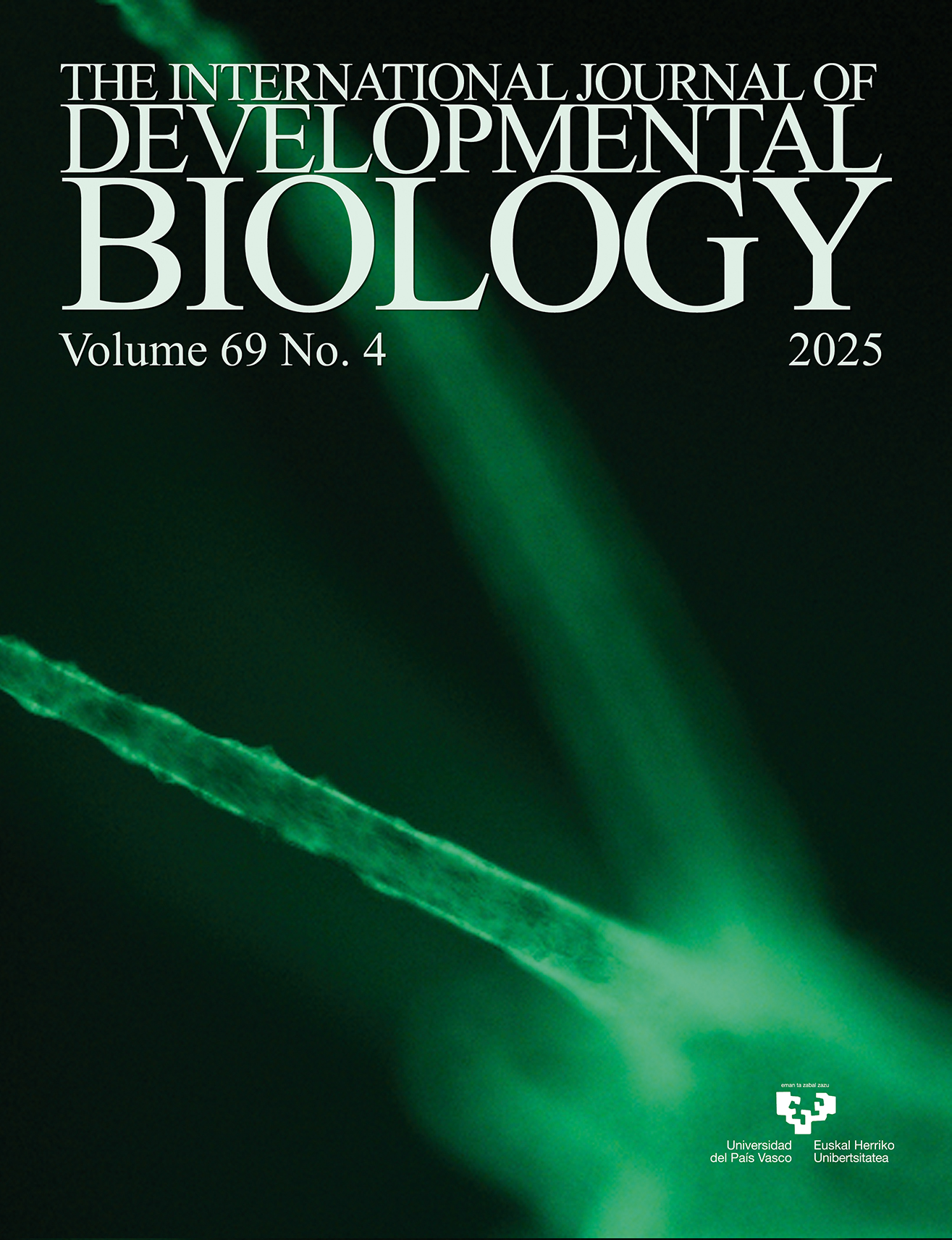
Nodular tentacles evoked by siRNA knockdown of Hv-fgf-c in Hydra vulgaris Traffic light. The GFP-transgenic tentacle ectoderm is shown 15 days post electroporation at 40x magnification. For further details, see the article by Ohler et al. in this issue, pp. 173-185.
OPEN ACCESS
Int. J. Dev. Biol. (2025) 69: 161-172
https://doi.org/10.1387/ijdb.250129km
ABSTRACT
Most major organs, like muscles, bones, vessels and kidneys, develop from the mesoderm, one of three germ cell layers in triploblastic organisms. Sialic acids significantly affect embryonic development by regulating cell division, migration and death through signaling pathways and cell adhesion, which support morphogenesis. Loss of early biosynthetic enzymes reduces embryonic viability and leads to complex phenotypes, while the loss of terminal enzymes primarily results in tissue-specific defects in mesoderm-derived organs. Key sialylated glycoproteins involved in the developmental processes of mesoderm and mesoderm-derived organs have been identified across various species as major effectors. These enzymes and glycoproteins are of significant interest and are discussed in the present review.
OPEN ACCESS
The Hydra FGF family – dispersed across the genome and expressed locally
Int. J. Dev. Biol. (2025) 69: 173-185
https://doi.org/10.1387/ijdb.250118mh
ABSTRACT
Although the nonbilaterian Hydra has a simple body architecture, its signaling systems are as complex as those of Bilateria. We here add data to the fibroblast growth factor signaling system which has previously been identified in Hydra as essential for bud detachment. The localized expression patterns of its, now fifteen, FGFs indicate additional, yet unidentified, functions in sometimes very small subsets of cells at the body termini and/or during budding or in testes. Presence of a typical signal sequence (prediction >93%) in only 30% of the Hydra FGFs suggests mechanisms of action (and/or secretion) other than the canonical ones. A knockdown approach using siRNA revealed a potential role for Hv-FGF-c in tentacle morphogenesis. Our results document a high complexity of FGF signaling sites in Hydra and open the field for a detailed analysis of their functions.
OPEN ACCESS
Int. J. Dev. Biol. (2025) 69: 187-194
https://doi.org/10.1387/ijdb.250101rm
ABSTRACT
Failure to properly regenerate and maintain tissues is a root cause of disease and aging. One goal of regenerative biology is to unveil mechanisms by which simpler model organisms successfully regenerate so that we can better understand how to control the cellular and molecular switches in human tissues. Planarians have become a favored model system for these studies because of their amazing capacity to fully and rapidly regenerate following amputation, but also for their response to other stressors such as starvation. Planarians undergo proportional degrowth when starved, a unique process that balances stem cell proliferation and apoptotic cell death, autophagy-mediated cell turnover, and energy sensing. We chose a pharmacological approach to study autophagy in planarians by using chloroquine, an autophagy inhibitor. We found that head regression and tissue lysis were induced by chloroquine in whole planarians and that starved planarians were more sensitive than their fed counterparts. While chloroquine treatment did not prevent tissue regeneration following injury, it caused regeneration delays and disruptions in photoreceptor replacement. Finally, we show that an...
OPEN ACCESS
Int. J. Dev. Biol. (2025) 69: 195-202
https://doi.org/10.1387/ijdb.250162rs
ABSTRACT
Dynein axonemal assembly factors (DNAAFs) play crucial roles in the formation and function of motile cilia, and their dysfunction often results in primary ciliary dyskinesia (PCD). We report the spatio-temporal expression patterns of dnaaf5 and dnaaf9 mRNA in zebrafish embryos, providing insight into their possible functions during development. We show that dnaaf5 and dnaaf9 mRNAs are expressed in motile ciliated tissues, such as the Kupffer’s vesicle, pronephros, floor plate, brain and olfactory placode. The dnaaf5 and dnaaf9 crispants develop ciliopathic defects during zebrafish development. These data suggest that dnaaf5 and dnaaf9 may regulate motile cilia biogenesis and function in zebrafish. Our findings suggest functional redundancy and divergence among dynein arm assembly factors in vertebrates.
OPEN ACCESS
Spatiotemporal dynamics of lineage-specific epithelial maturation in the developing mouse stomach
Int. J. Dev. Biol. (2025) 69: 203-215
https://doi.org/10.1387/ijdb.250171ht
ABSTRACT
At birth, the gastric epithelium is immature and progressively matures during postnatal development to establish adult tissue architecture and function. Although this process has been investigated at functional, proteomic and transcriptomic levels, the precise temporal and spatial dynamics by which individual epithelial lineages initiate differentiation, exit proliferative states and establish adult homeostasis remain incompletely understood. Here, we performed a comprehensive immunofluorescence analysis of the mouse stomach from mid-gestation through adulthood, examining fetal markers, lineage-specific functional epithelial markers, and the proliferation marker KI67. By integrating functional epithelial marker expression with proliferative status, we delineated distinct maturation trajectories for pit, parietal, and chief cell lineages. Notably, parietal and chief cells exhibited a robust proliferative phase from late gestation to early postnatal stages, followed by a marked decline in proliferative activity, whereas pit cells showed the emergence of proliferative progenitors primarily during later postnatal stages. Moreover, we identified region-specific differences in the...in situ atlas of gastric epithelial maturation and provides a framework for understanding lineage- and region- specific mechanisms governing postnatal gastric development.