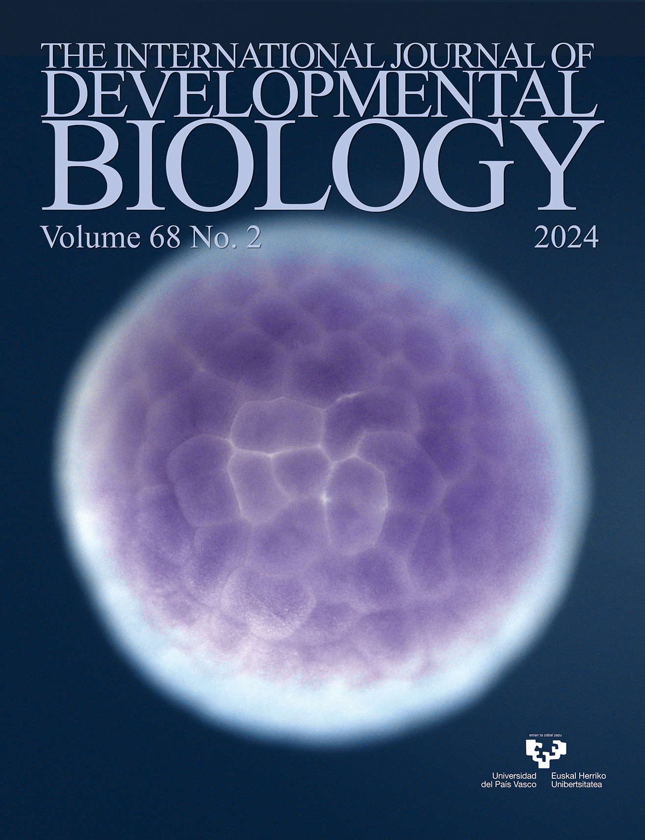
Whole-mount in situ hybridization showing thg1l expression (purple staining) in the animal blastomeres of a Xenopus laevis stage 7 blastula. For further details, see the article by Martini et al. in this issue, pp. 85-91.
OPEN ACCESS
Coenocystic oogenesis - modification of or deviation from the germ cell cyst paradigm?
Int. J. Dev. Biol. (2024) 68: 47-53
https://doi.org/10.1387/ijdb.240064mk
ABSTRACT
Invertebrate and vertebrate species have many unusual cellular structures, such as long- or short-lived cell-in-cell structures and coenocytes. Coenocytes (often incorrectly described as syncytia) are multinuclear cells derived, unlike syncytia, not from the fusion of multiple cells but from multiple nuclear divisions without cytokinesis. An example of a somatic coenocyte is the coenocytic blastoderm in Drosophila. An astonishing property of coenocytes is the ability to differentiate the nuclei sharing a common cytoplasm into different subpopulations with different fate trajectories. An example of a germline coenocyte is the oogenic precursor of appendicularian tunicates, which shares many features with the somatic coenocyte of Drosophila. The germline coenocyte (coenocyst) is quite an unexpected structure because in most animals, including Drosophila, Xenopus, and mice, oogenesis proceeds within a group (cyst, nest) of sibling cells (cystocytes) connected by the intercellular bridges (ring canals, RCs) derived from multiple divisions with incomplete cytokinesis of a progenitor cell called the cystoblast. Here, I discuss the differences and similarities between cystocyte-based and...Drosophila. I also describe cell-in-cell structures that although not mechanistically, cytologically, or molecularly connected to somatic or germline coenocytes, are both unorthodox and intriguing cytological phenomena rarely covered by scientific literature.
OPEN ACCESS
TGF-β signaling molecules in Hydra: role of BMP and BMP inhibitors during pattern formation
Int. J. Dev. Biol. (2024) 68: 55-64
https://doi.org/10.1387/ijdb.240009sg
ABSTRACT
Understanding the evolution of body plans has been one of the major areas of investigation in developmental and evolutionary biology. Cnidaria, the sister group to bilaterians, provides an opportunity to elucidate the origin and evolution of body axes. Hydra, a freshwater cnidarian, is a useful model to study signaling pathways governing pattern formation, which are conserved up to vertebrates including humans. The transforming growth factor β (TGF-β) signaling pathway is one of the fundamental pathways that regulate axis formation and organogenesis during embryonic development. In this article, we discuss the TGF-β pathway members identified in Hydra along with other cnidarians with an emphasis on bone morphogenetic proteins (BMPs) and their inhibitors. TGF-β members, especially those involved in BMP signaling pathway, are mainly involved in maintaining the Organizer region and patterning the body axis in Hydra. Identification of other members of this pathway in Hydra and fellow cnidarians would provide insights into the evolution of body axes and pattern formation in more complex metazoans.
OPEN ACCESS
Molecular signaling directing neural plate border formation
Int. J. Dev. Biol. (2024) 68: 65-78
https://doi.org/10.1387/ijdb.230231me
ABSTRACT
During embryonic development, the vertebrate embryonic epiblast is divided into two parts including neural and superficial ectoderm. The neural plate border (NPB) is a narrow transitional area which locates between these parts and contains multipotent progenitor cells. Despite its small size, the cellular heterogeneity in this region produces specific differentiated cells. Signaling pathways, transcription factors, and the expression/repression of certain genes are directly involved in these differentiation processes. Different factors such as the Wnt signaling cascade, fibroblast growth factor (FGF), bone morphogenetic protein (BMP) signaling, and Notch, which are involved in various stages of the growth, proliferation, and differentiation of embryonic cells, are also involved in the determination and differentiation of neural plate border stem cells. Therefore, it is essential to consider the interactions and temporospatial coordination related to cells, tissues, and adjacent structures. This review examines our present knowledge of the formation of the neural plate border and emphasizes the requirement for interaction between different signaling pathways, including the BMP and...
OPEN ACCESS
Int. J. Dev. Biol. (2024) 68: 79-83
https://doi.org/10.1387/ijdb.240060jd
ABSTRACT
Mutations in the gene encoding Tre2/Bub2/Cdc16 (TBC)1 domain family member 24 (TBC1D24) protein are associated with a variety of neurological disorders, ranging from non-syndromic hearing loss to drug-resistant lethal epileptic encephalopathy and DOORS syndrome [Deafness, Onychodystrophy, Osteodystrophy, intellectual disability (formerly referred to as mental Retardation), and Seizures]. TBC1D24 is a vesicle-associated protein involved in neural crest cell and neuronal migration, maturation, and neurotransmission. In the cochlea, TBC1D24 has been detected in auditory neurons, but few reliable and convergent data exist about the sensory epithelium. Here, the expression of TBC1D24 has been characterized via immunolabelling throughout the postnatal maturation of the mouse cochlear sensory epithelium. TBC1D24 was detected in glia-like non-sensory epithelial cells during early developmental stages. In contrast, TBC1D24 was virtually absent in adjacent sensory hair cells. This expression distinguishing non-sensory from sensory epithelial cells almost disappears around the onset of hearing. Until now, TBC1D24 was mainly described as a neuronal protein either in the brain or in the...
OPEN ACCESS
Expression analysis of thg1l during Xenopus laevis development
Int. J. Dev. Biol. (2024) 68: 85-91
https://doi.org/10.1387/ijdb.240033ma
ABSTRACT
The tRNA-histidine guanylyltransferase 1-like (THG1L), also known as induced in high glucose-1 (IHG-1), encodes for an essential mitochondria-associated protein highly conserved throughout evolution, that catalyses the 3′–5′ addition of a guanine to the 5′-end of tRNA-histidine (tRNAHis). Previous data indicated that THG1L plays a crucial role in the regulation of mitochondrial biogenesis and dynamics, in ATP production, and is critically involved in the modulation of apoptosis, cell-cycle progression and survival, as well as in cellular stress responses and redox homeostasis. Dysregulations of THG1L expression play a central role in various pathologies, including nephropathies, and neurodevelopmental disorders often characterized by developmental delay and cerebellar ataxia. Despite the essential role of THG1L, little is known about its expression during vertebrate development. Herein, we examined the detailed spatio-temporal expression of this gene in the developing Xenopus laevis. Our results show that thg1l is maternally inherited and its temporal expression suggests a role during the earliest stages of embryogenesis. Spatially, thg1l mRNA localizes in the ectoderm and...thg1l transcripts mostly localise in neural crests and their derivatives, somites, developing kidney and central nervous system, therefore largely coinciding with territories displaying intense energy metabolism during organogenesis in Xenopus.