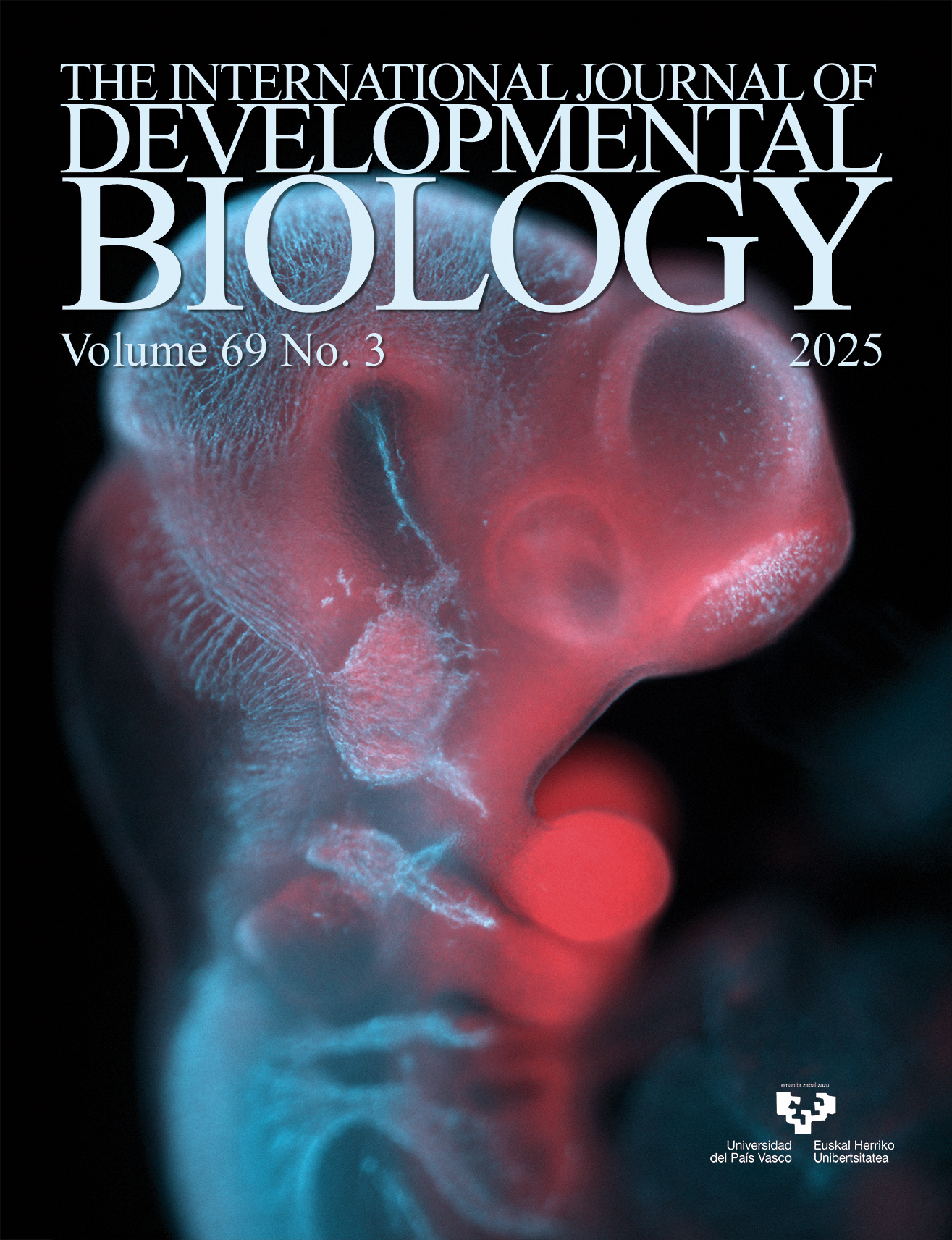
TuJ1/Tubb3 immunostaining (blue) and Wnt1Cre2; Rosa26rtdTomato/tdTomato (red) labeling of neural crest cells and their derivatives in an E10.5 murine embryo, exploring the role of Mllt11 in neural crest cell development and masseter innervation. For further details, see the article by Zinck et al. in this issue, pp. 123-131.
OPEN ACCESS
Int. J. Dev. Biol. (2025) 69: 109-122
https://doi.org/10.1387/ijdb.250018mp
ABSTRACT
How the dorsal thalamus of amniotes (reptiles, birds, and mammals) is organized remains an important but incompletely answered question. Identification of meaningful subdivisions would greatly aid in its understanding. Because the dorsal thalamus is more simply organized during development, studies have examined this structure during embryogenesis. Most reports using this approach have examined the developing dorsal thalamus in mammals and birds. Only rarely has the development of the dorsal thalamus been investigated in reptiles. Regardless, any approach to identify subdivisions, the presumed building blocks of the dorsal thalamus, should include representatives of all three classes of vertebrates. To fill this gap in knowledge, the development of the dorsal thalamus was investigated in Alligator mississippiensis, a member of the reptilian group most closely related to birds. As the first detailed study of its kind, cytoarchitecture and calretinin expression were used to examine dorsal thalamus development. Three subdivisions, termed tiers, and the individual nuclei originating from each tier, were identified. These three tiers were similar to the subdivisions found in birds and,...
OPEN ACCESS
Int. J. Dev. Biol. (2025) 69: 123-131
https://doi.org/10.1387/ijdb.240249ai
ABSTRACT
The development of cranial nerves, including the trigeminal nerve, and the formation of neuromuscular junctions (NMJs) are crucial processes for craniofacial motor function. Mllt11/Af1q/Tcf7c (hereafter Mllt11), a novel type of cytoskeletal-interacting protein, has been implicated in neuronal migration and neuritogenesis during central nervous system development. However, its role in peripheral nerve development and NMJ formation remains poorly understood. This study investigates the function of Mllt11 during trigeminal ganglion development and its impact on motor innervation of the masseter muscle. We report Mllt11 expression in the developing trigeminal ganglia, suggesting a potential role in cranial nerve development. Using a conditional knockout mouse model to delete Mllt11 in Wnt1-expressing neural crest cells, we assessed trigeminal ganglion development and innervation of the masseter muscle in the jaw. Surprisingly, we found that Mllt11 loss does not affect the initial formation of the trigeminal ganglion but disrupts its placodal vs. neural crest cellular composition. Furthermore, we showed that conditional inactivation of Mllt11 using Wnt1Cre2 led to a reduction of...+ branchiomotor neurons in rhombomere 2, indicating altered trigeminal motor innervation. This was due to the surprising finding that the Wnt1Cre2/+ mouse driver promoted aberrant recombination and reporter gene expression within branchiomotor neuron pools in rhombomere 2, as well as targeting neural crest cell populations. Our findings show that Mllt11 regulates the cellular composition of the trigeminal ganglion and is essential for proper trigeminal motor innervation in the masseter muscle.
OPEN ACCESS
Int. J. Dev. Biol. (2025) 69: 133-142
https://doi.org/10.1387/ijdb.250065jr
ABSTRACT
Interactions between ephrins and their Eph receptors regulate a broad range of cellular processes, including attraction, repulsion, adhesion and migration, all of which play crucial roles in tissue remodeling and homeostasis. While several ephrin ligands and Eph receptors are known to be expressed in the developing kidney, their specific roles, particularly during nephrogenesis, remain poorly understood. The development of the Xenopus pronephros provides an accessible and relatively simple model for studying vertebrate nephrogenesis. Through a comprehensive gene expression analysis of all ephrin ligands and Eph receptors present in Xenopus genomes, we have identified members of the Eph-ephrin signaling pathway that may contribute to pronephric development. Among them, efna3, which encodes an ephrin ligand, is strongly expressed in the ventral region of the pronephric anlage and later in the intermediate and distal segments of the developing tubule. This expression pattern is strikingly complementary to that of epha4 and epha7, which are expressed in the region forming the proximal tubule. This suggests a potential role for efna3-epha4/epha7 signaling in establishing a boundary...
OPEN ACCESS
Int. J. Dev. Biol. (2025) 69: 143-150
https://doi.org/10.1387/ijdb.250046yh
ABSTRACT
Carnitine palmitoyltransferase 1 (CPT1) is a key regulatory enzyme in fatty acid metabolism, responsible for the translocation of long-chain fatty acids into the mitochondria for β-oxidation in diverse biological contexts. Recent studies implicated the critical role of cpt1 genes during zebrafish development and heart regeneration; however, a comprehensive characterization of their spatiotemporal expression dynamics remains lacking. Here, we systematically analyzed the expression profiles of four cpt1 paralogs (cpt1aa, cpt1ab, cpt1b, and cpt1a2b) during zebrafish embryogenesis and the expression of cpt1ab and cpt1b during zebrafish heart regeneration. Our results reveal that these paralogs exhibit distinct spatiotemporal expression patterns during zygotic development. While cpt1aa and cpt1ab share high sequence conservation (77%), their expression patterns diverge substantially. Conversely, cpt1ab and cpt1b display convergent cardiac and somitic expression despite lower sequence similarity (53%). Following ventricular ablation, cpt1b expression transiently ceased then recovered during regeneration, whereas cpt1ab remained unchanged. These findings shed light on the evolutionary...cpt1 paralogs, which establish a critical foundation for elucidating paralog-specific roles in fatty acid metabolism during vertebrate development and regeneration.
OPEN ACCESS
Int. J. Dev. Biol. (2025) 69: 151-158
https://doi.org/10.1387/ijdb.250093mt
ABSTRACT
The skull of amniotes is categorized into three conditions based on skeletal arrangement in the temporal region: anapsid, synapsid and diapsid. Mammals (class Mammalia), a descendent lineage of the clade Synapsida, possess the synapsid skull, which is characterized by a single lower temporal arch that ventrally borders the lower temporal fenestra. Although we previously suggested, based on the data from placental mammals, that the reduction in the expression domain of the upstream osteogenic genes Msx2 and Runx2 in the embryonic temporal mesenchyme might have played a role in the evolution of synapsid skulls, the molecular basis of synapsid skull evolution is still largely unknown. In this study, we investigated expression patterns of four osteogenic genes (two upstream genes Msx2 and Runx2 and two downstream genes Sp7 and Sparc) in the embryonic and neonatal temporal region of the gray short-tailed opossum Monodelphis domestica, the most commonly used experimental marsupial model, in order to more thoroughly understand the molecular basis of development of synapsid skulls unique to mammals. We found that M. domestica embryos and neonates display very restricted expressions of Msx2 and/or Runx2 in the dermal bone precursors in the temporal region, as two placental species do (the house mouse Mus musculus and the greater horseshoe bat Rhinolophus ferrumequinum). Spatially restricted expression of Msx2 and Runx2 in the embryonic temporal region may be a foundation for creating the "advanced" synapsid skull shared by all mammals where only three dermal bones configure the temporal region.
OPEN ACCESS
Int. J. Dev. Biol. (2025) 69: 159-159
https://doi.org/10.1387/ijdb.250154sb
ABSTRACT
Following publication of this article, the authors identified typographical errors in the primer sequences listed in Table 1 in the Materials and Methods section. The corrected table is published.