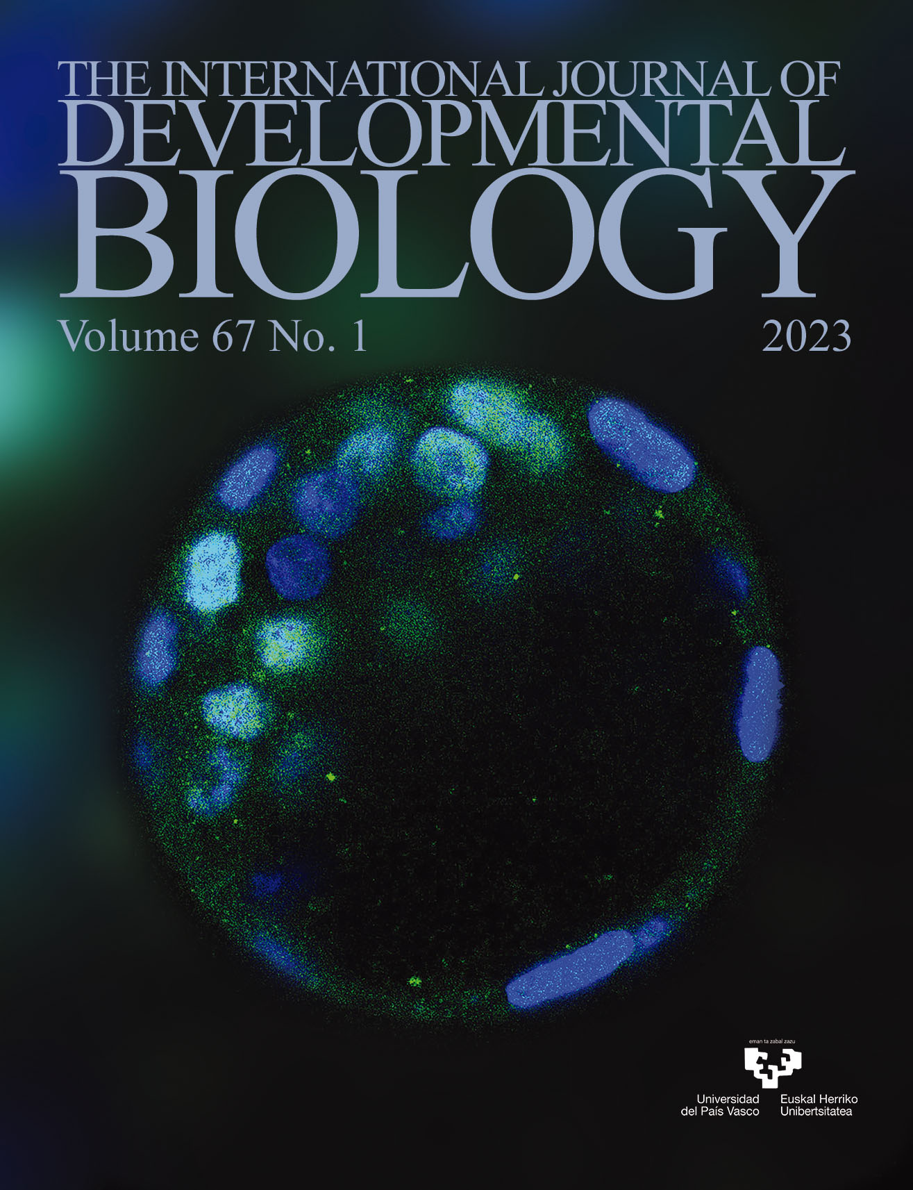
Nanog expression in mouse blastocyst. Green signals indicate Nanog protein located in the nuclei of inner cell mass, blue signals (with Hoechst 33342) show the cell nuclei. For further details, see the article by Fatma Uysal et al. in this issue, pp. 1-8.
OPEN ACCESS
DNA methyltransferase (Dnmt) silencing causes increased Cdx2 and Nanog levels in surviving embryos
Int. J. Dev. Biol. (2023) 67: 1-8
https://doi.org/10.1387/ijdb.230040oc
ABSTRACT
Epigenetic mechanisms are one of the essential regulators of gene expression which do not involve altering the primary nucleotide sequence. DNA methylation is considered among the most prominent epigenetic mechanisms in controlling the functions of genes related to cell differentiation, cell cycle, cell survival, autophagy, and embryo development. DNA methyl transferases (Dnmts) control DNA methylation, the levels of which are differentially altered during embryonic development, and may determine cell differentiation fate as in the case of pluripotent inner cell mass (ICM) or trophectoderm (TE). In this study, we aimed to analyze the role of Dnmt1 and Dnmt3a enzymes in ICM (using the Nanog marker) and TE (using the Cdx2 marker) differentiation, autophagy (using p62 marker), reactive oxygen species (ROS) production, and apoptosis (using TUNEL) during mouse preimplantation embryo development. Following knockdown of Dnmt1 and Dnmt3a in zygotes, expression levels of Cdx2 in the trophectoderm and Nanog in the inner cell mass were measured, as well as p62 levels, reactive oxygen species (ROS) production, and apoptosis levels after 96 hours in embryo culture. We found that knockdown of...
Int. J. Dev. Biol. (2023) 67: 9-17
https://doi.org/10.1387/ijdb.220150jl
ABSTRACT
Acute myocardial infarction (AMI) is myocardial necrosis caused by the complete or partial obstruction of a coronary artery. Circular RNAs (circRNAs) have been proven as regulators in the progression of various human diseases, including AMI. However, the role of novel circ-JA760602 in AMI remains unknown. Here, we investigated the role of circ-JA760602 in modulating the apoptosis of hypoxia-induced AMI cells using the AC16 cardiomyocyte in vitro cell model. The expression of circ-JA760602 in AC16 cardiomyocytes subjected to hypoxia was measured by quantitative real-time polymerase chain reaction (qRT-PCR). Cell viability was measured by cell counting kit-8 (CCK-8) assay. Apoptosis of cardiomyocytes was evaluated by TUNEL assay and flow cytometry analysis. The cellular location of circ-JA760602 was identified through fluorescence in situ hybridization (FISH) assay and subcellular fractionation assay. The downstream molecular mechanisms of circ-JA760602 were demonstrated by luciferase reporter assay, RNA binding protein immunoprecipitation (RIP) assay and chromatin immunoprecipitation (ChIP) assay. Rescue assays were performed to demonstrate the effects of BCL2 knockdown on...
Int. J. Dev. Biol. (2023) 67: 19-25
https://doi.org/10.1387/ijdb.220173db
ABSTRACT
SOX transcription factors play key roles in cell differentiation and cell fate determination during development. Using single-cell RNA-sequencing data, we examined the expression profiles of Sox genes in the mouse incisor dental pulp. Our analysis showed that Sox4, Sox5, Sox9, Sox11, and Sox12 are mainly expressed in mesenchymal stem/stromal cells (MSCs) representing osteogenic cells at different stages of differentiation. We found that in several MSCs, Sox genes co-expressed with regulatory genes such as Sp7, Satb2, Msx1, Snai2, Dlx1, Twist2, and Tfap2a. In addition, Sox family genes colocalized with Runx2 and Lef1, which are highly enriched in MSCs undergoing osteoblast differentiation. A protein interaction network analysis uncovered that CREBBP, CEBPB, TLE1, TWIST1, and members of the HDAC and SMAD families are interacting partners of RUNX2 and LEF1 during skeletal development. Collectively, the distinct expression patterns of the SOX transcription factors suggest that they play essential regulatory roles in directing lineage-specific gene expression during differentiation of MSCs.