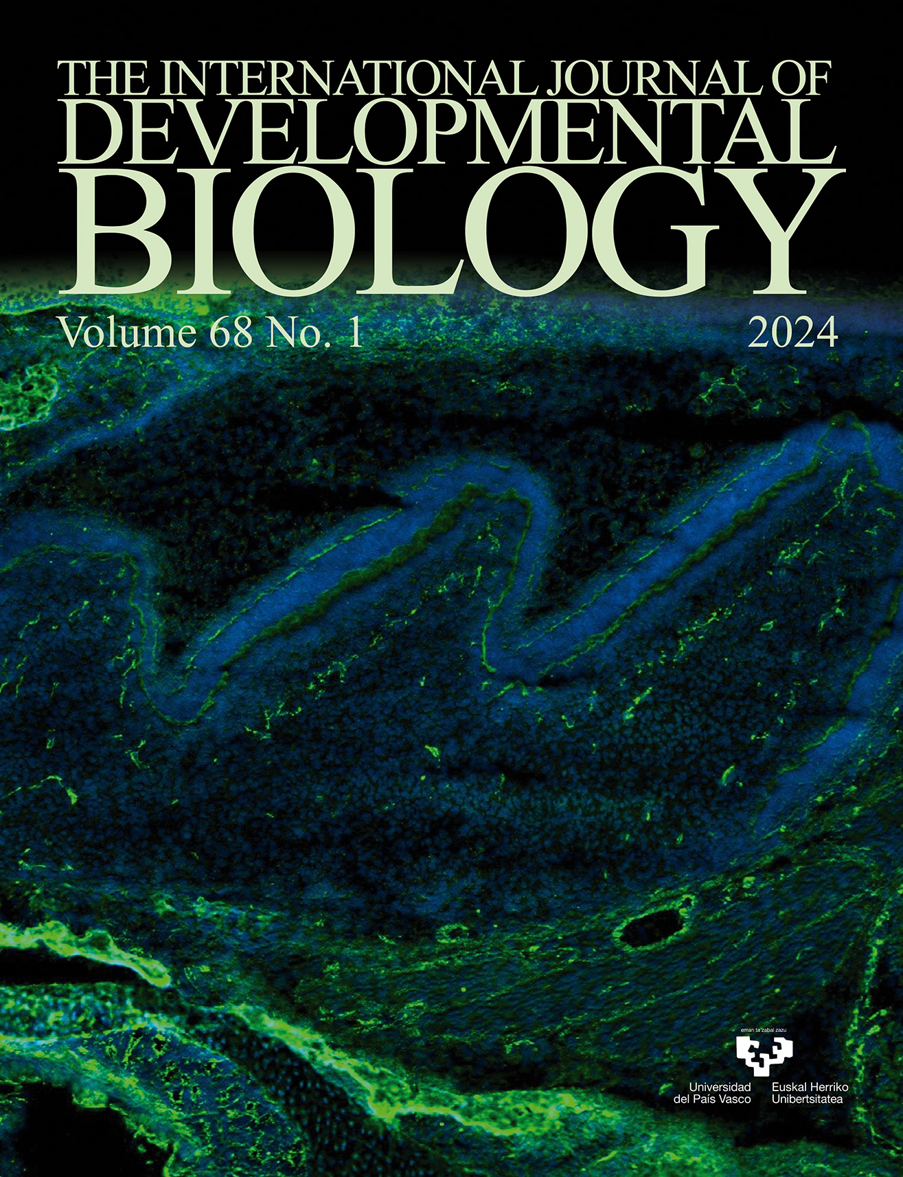
Immunofluorescence showing expression of Zonula Occludens 1 (in green) and DAPI (in blue) in embryonic day 19.5 (E19.5) mouse molars. For further details, see the article by Jiménez-Rojo et al. in this issue, pp. 19-24.
OPEN ACCESS
Estrogen signaling in development: recent insights from the zebrafish
Int. J. Dev. Biol. (2024) 68: 1-7
https://doi.org/10.1387/ijdb.230116rw
ABSTRACT
While traditionally recognized as a sex hormone, estrogen has a potent effect on the development of tissues beyond those of the reproductive system. Estrogen synthesis enzymes and estrogen receptors are broadly expressed in vertebrate tissues, further indicating their importance in various processes. These include the tissues of the zebrafish, which is a particularly suitable model for studying early development due to its rapid ex utero ontogeny and conserved genetic and cellular composition with other vertebrates. In this review, we provide readers with an overview of estrogen signaling, discuss important attributes of the zebrafish animal model with a special focus on the kidney, and explore recent insights from zebrafish studies about the roles of estrogen signaling in organogenesis across germ layer derivatives that range from the kidney to the brain and liver.
OPEN ACCESS
Understanding megasporogenesis through model plants: contemporary evidence and future insights
Int. J. Dev. Biol. (2024) 68: 9-17
https://doi.org/10.1387/ijdb.230222mk
ABSTRACT
The megasporangium serves as a model system for understanding the concept of individual cell identity, and cell-to-cell communication in angiosperms. As development of the ovule progresses, three distinct layers, the epidermal (L1), the subepidermal or the hypodermal (L2) and the innermost layers (L3) are formed along the MMC (megaspore mother cell). The MMC, which is the primary female germline cell, is initiated as a single subepidermal cell amongst several somatic cells. MMC development is governed by various regulatory pathways involving intercellular signaling, small RNAs and DNA methylation. The programming and reprograming of a single nucellar cell to enter meiosis is governed by ‘permissive’ interacting processes and factors. Concomitantly, several nucellar sister cells are prevented from germline fate also by a set of ‘repressive’ factors. However, in certain angiosperms, anomalies in development of the female gametophyte have been observed. The sporophytic tissue surrounding the female gametophyte affects the gametophyte in multiple ways. The role of genes and transcription factors in the development of the MMC and in the regulation of various processes studied...Arabidopsis is explained in detail in this paper. However, as angiosperms display enormous diversity, it is important to investigate early stages of megasporogenesis in other plant systems as well. Such studies provide valuable insights in understanding the regulation of megasporogenesis and the evolution of the female gametophyte from gymnosperms to flowering plants.
OPEN ACCESS
Disrupted odontoblast differentiation and dentin dysplasia in Epiprofin-deficient mice
Int. J. Dev. Biol. (2024) 68: 19-24
https://doi.org/10.1387/ijdb.240029lj
ABSTRACT
Tooth formation is a process tightly regulated by reciprocal interactions between epithelial and mesenchymal tissues. These epithelial-mesenchyme interactions regulate the expression of target genes via transcription factors. Among the regulatory elements governing this process, Epiprofin/Sp6 is a zinc finger transcription factor which is expressed in the embryonic dental epithelium and in differentiating pre-odontoblasts. Epiprofin knockout (Epfn-/-) mice present severe dental abnormalities, such as supernumerary teeth and enamel hypoplasia. Here, we describe dentin defects in molars and incisors of Epfn-/- mice. We observed that in the absence of Epfn, markers of early odontoblast differentiation, such as alkaline phosphatase activity, Dsp/Dpp expression, and Collagen Type I deposition, are downregulated. In addition, the expression of tight and gap junction proteins was severely impaired in the predontoblastic cell layer of developing Epfn-/- molars. Altogether, our data shows that Epfn is crucial for the proper differentiation of dental mesenchymal cells towards functional odontoblasts and subsequent dentin-matrix deposition.
OPEN ACCESS
Enhancement of neural crest formation by mechanical force in Xenopus development
Int. J. Dev. Biol. (2024) 68: 25-37
https://doi.org/10.1387/ijdb.230273tm
ABSTRACT
In vertebrate development, ectoderm is specified into neural plate (NP), neural plate border (NPB), and epidermis. Although such patterning is thought to be achieved by molecular concentration gradients, it has been revealed, mainly by in vitro analysis, that mechanical force can regulate cell specification. During in vivo patterning, cells deform and migrate, and this applies force to surrounding tissues, shaping the embryo. However, the role of mechanical force for cell specification in vivo is largely unknown. In this study, with an aspiration assay and atomic force microscopy, we have demonstrated that tension on ectodermal cells decreases laterally from the midline in Xenopus early neurula. Ectopically applied force laterally expanded the neural crest (NC) region, a derivative of the NPB, whereas force relaxation suppressed it. Furthermore, force application activated both the FGF and Wnt pathways, which are required for NC formation during neuroectodermal patterning. Taken together, mechanical force is necessary for NC formation in order to regulate signaling pathways. Furthermore, molecular signals specify the NP and generate force on neighboring tissue, the NPB, with its...
OPEN ACCESS
Developmental relationship between junctional epithelium and epithelial rests of Malassez
Int. J. Dev. Biol. (2024) 68: 39-45
https://doi.org/10.1387/ijdb.230243sl
ABSTRACT
Keratin 17 (K17) is thought to be a candidate target gene for regulation by Lymphoid Enhancer Factor-1 (Lef-1). K17 is a marker that distinguishes junctional epithelium (JE) from epithelial rests of Malassez (ERM). However, the relationship of Lef-1 to K17 is not clear in this context. Moreover, the expression of other keratins such as K5, K6, K7 and K16 is not reported. Therefore, the aim of our study was to assay the expression of K5, K6, K7, K14, K16, K17 and Lef-1 in postnatal developing teeth, and clarify the corresponding immunophenotypes of the JE and ERM. Upper jaws of Wistar rats aged from postnatal (PN) day 3.5 to PN21 were used and processed for immunohistochemistry. K5 and K14 were intensely expressed in inner enamel epithelium (IEE), reduced enamel epithelium (REE), ERM and JE. There was no staining for K16 in the tissue, except for strong staining in the oral epithelium. Specifically, at PN3.5 and PN7, K17 was initially strongly expressed and then negative in the IEE. At PN16 and PN21, both REE and ERM were strongly stained for K17, whereas K17 was negative in the JE. In addition, K6, K7 and Lef-1 were not detected in any tissue investigated. REE and ERM have an...