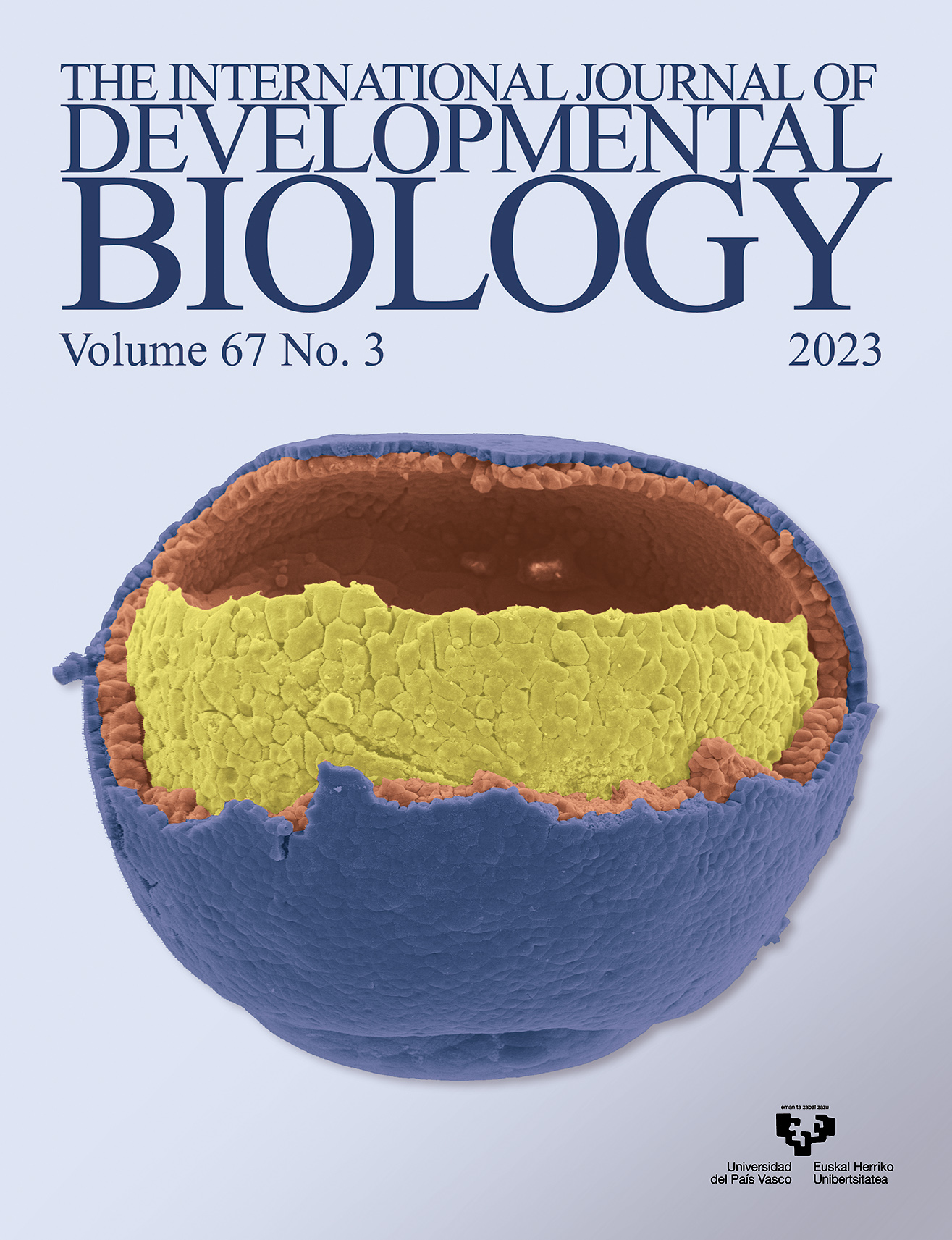
Removing part of the ectoderm (blue, outer epithelial layer; orange, deep layer) exposes the mesendoderm (yellow), a gut- and blood-forming tissue primordium in the frog early embryo. An intricate pattern of cell-cell adhesion and repulsion ensures its directional movement upward to the animal pole (top). For further details, see the article by Nagel and Winklbauer in this issue, pp. 79-90.
Insights into the role of the Wnt signaling pathway in the regeneration of animal model systems
Int. J. Dev. Biol. (2023) 67: 65-78
https://doi.org/10.1387/ijdb.220144yl
ABSTRACT
Regeneration enables the regrowth and restoration of missing body parts. It is a common phenomenon among animals. However, only some species exhibit remarkable regeneration capabilities and can regenerate organs such as limbs, lenses or hearts. Regeneration has been widely studied, thereby giving rise to new fields, such as regenerative medicine. Furthermore, regeneration has the potential to be applied to the human body. However, the molecular mechanisms governing this process should be elucidated first. Recent advancements in research methods have led to the identification of numerous signaling pathways involved in regeneration. One of them, the Wnt transduction pathway, is an ancient and evolutionarily conserved pathway that plays an important role in both embryonic development and regeneration. The Wnt pathway plays an important role during the regeneration process, as it is implicated in cell fate determination, cell migration, cell polarity and adult cell homeostasis. To date, two major Wnt pathways have been identified: the canonical (β-catenin dependent) pathway and the non-canonical pathway. The latter pathway can be further divided into planar cell polarity, the...
OPEN ACCESS
Polarized contact behavior in directionally migrating Xenopus gastrula mesendoderm
Int. J. Dev. Biol. (2023) 67: 79-90
https://doi.org/10.1387/ijdb.230123rw
ABSTRACT
The control of cell-cell adhesion and detachment is essential for collective migration and cell rearrangement. Here, we have used the contact behavior of Xenopus gastrula mesoderm explants migrating directionally on ectoderm conditioned substratum to study the regulation of active cell-cell detachment. When colliding laterally, explants repelled each other, whereas they fused front-to-back when aligned in the direction of migration. For this mesoderm polarization by the substratum, we identified three control modules. First, PDGF-A signaling normally suppresses contact-induced collapse of lamellipodia in a polarized manner. Disruption of PDGF-A function, or of Xwnt6, decreased the polarization of explant contact behavior. Second, the Wnt receptor Xfz7 acted upstream of the kinase Pak1 to control explant fusion independently of PDGF-A-promoted lamellipodia stability. Third, ephrinB1 acted with Dishevelled (Dvl) in front-to-back explant fusion. The second and third modules have been identified previously as regulators of tissue separation at the ectoderm-mesoderm boundary. On non-polarizing, fibronectin-coated substratum, they controlled repulsion between explants in the same way as...
Int. J. Dev. Biol. (2023) 67: 91-100
https://doi.org/10.1387/ijdb.230156sa
ABSTRACT
Although preterm birth is among the preventable causes of maternal and infant death, its mechanism has not yet been clarified. When evaluated in terms of the results, the psycho-social burden of mother-infant losses and the costs of rehabilitation, care, and treatment for postpartum sequelae are high. When evaluated in terms of its causes, infection/inflammation has an important place. Therefore, it is essential to understand the role of pro- and anti-inflammatory proteins in the process. In our study, apelin and apelin receptor (APJ) expression in the cervix-uterus and placental axis were evaluated at tissue and protein levels in pregnant and non-pregnant control, sham, PBS, and LPS groups in the infection model in which LPS induction was performed by midline laparotomy, in CD-1 mice. The evaluation of this axis regarding apelin and apelin receptor in the preterm birth model is new in the literature. Apelin is expressed more intensely in uterine epithelial cells than in the cervix. In the placenta, expression is more intense in the junctional zone compared to other zones. Apelin protein levels decrease significantly in the cervix and placenta whereas it increases in the uterus....
Int. J. Dev. Biol. (2023) 67: 101-108
https://doi.org/10.1387/ijdb.230154ng
ABSTRACT
Valproic acid (VPA), a neuroprotective agent and inhibitor of GSK3-β, along with human Adipose-Derived Stem Cells (hADSCs) have been proposed to be potential therapeutic agents for neurodegenerative disorders. In the present study, we have assessed the effects of VPA alone or in combination with hADSCs on oligodendrocyte differentiation, remyelination, and functional recovery in a mouse model of Multiple Sclerosis (MS). These MS-model mice were randomly divided into cuprizone, sham, VPA, hADSC, and VPA/hADSC groups, with 10 mice considered a control group (healthy mice). The hanging wire test was used to measure motor behavior. To estimate the level of myelination, we performed toluidine blue staining and immunofluorescent staining for OLIG2 and MOG-positive cells. Real-time PCR was used to evaluate the expression of β-catenin, human and mouse Mbp, Mog, and Olig2 genes. Remyelination and motor function improved in mice receiving VPA, hADSCs, and especially VPA/hADSCs compared to the Cup and Sham groups (P ). Additionally, the number of MOG and OLIG2 positive cells significantly increased in the VPA/hADSCs group compared to the Cup and Sham groups (P ). The expression of...