Int. J. Dev. Biol. 67: 109 - 114 (2023)
Developmental Biology: from genes to functional organism – news from the 3rd Meeting of the Visegrád Group Society for Developmental Biology
Open Access | Meeting Report | Published: 28 December 2023
Abstract
The third meeting of the Visegrád Group Society for Developmental Biology (V4SDB) was held on September 8th-10th, 2023 in Warsaw, Poland. It was a continuation of previous meetings, the first organized in the Czech Republic in 2018 and the second in Hungary in 2021. Similarly to the previous meetings, the organizers created a friendly platform for networking and science sharing. The conference gathered an excellent group of 160 researchers working on various animal models, who during lecture and poster sessions discussed a broad range of subjects, including early embryonic development, organogenesis, genetic and epigenetic control of developmental processes, stem cells and regeneration, cellular dynamics and migration in developmental biology, and in vitro models in development and disease. Additionally, two satellite events were organized: the Young Developmental Biologists’ Forum, which gave young researchers an opportunity to share and promote their work and to participate in hands-on courses, and an outreach initiative “Developmental Biology for Everyone”, which presented different aspects of developmental biology to a broad audience.
Keywords
developmental biology, organogenesis, early embryonic development, gametes, stem cells, regeneration, in vitro models, V4SDB
Introduction
The Visegrád Group Society for Developmental Biology (V4SDB) was established in 2018 to foster more efficient and integrated research in developmental biology across Central Europe, particularly in V4 countries: Czech Republic, Hungary, Poland and Slovakia. Although the majority of V4SDB members recruit from V4 residents, the society is open to members from other countries. The main goal of the V4SDB is to facilitate the exchange of knowledge and experimental methodology and to promote cooperation between the members.
The V4SDB community meets every 2 years in different V4 countries. On September 8th-10th 2023, we gathered in the beautiful and bustling city of Warsaw, the capital of Poland, where we were hosted by the Faculty of Biology, University of Warsaw (Figs. 1, 2A). The conference was co-organized by the Institute of Genetics and Animal Biotechnology, Polish Academy of Sciences, represented by Anna Piliszek, and the Faculty of Biology, University of Warsaw, represented by Anna Ajduk. With the support of other members of the V4SDB steering committee, Alexander W. Bruce (University of South Bohemia, Czech Republic), Vítêzslav Bryja (Masaryk University, Czech Republic), Marcela Buchtová (Masaryk University and Institute of Animal Physiology and Genetics, Czech Academy of Sciences, Czech Republic), Dušan Fabian (Institute of Animal Physiology, Slovak Academy of Sciences, Slovakia), Máté Varga (Eötvös Loránd University, Hungary), and Péter Vilmos (Biological Research Center, Hungarian Academy of Sciences, Hungary), Annas put together an exciting scientific program covering a broad spectrum of developmental biology topics, including early embryonic development, organogenesis, genetic and epigenetic control of developmental processes, stem cells and regeneration, cellular dynamics and migration in developmental biology, and in vitro models in development and disease (Fig. 1). Results presented in lectures, short talks, and posters came from a variety of animal models, both vertebrate and invertebrate (Fig. 2 D-F).
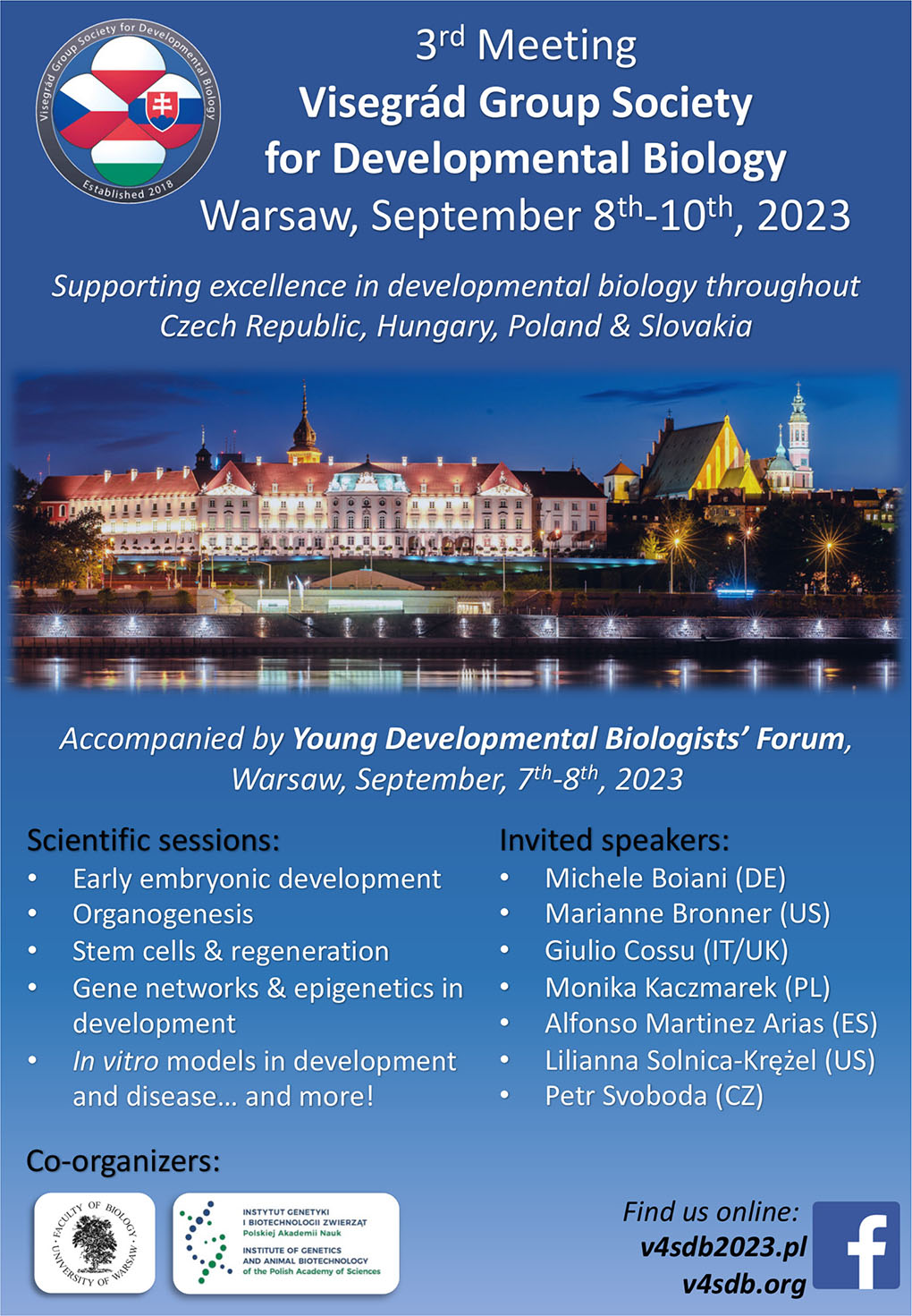
Fig. 1. Leaflet of the 3rd Visegrád Group Society for Developmental Biology (V4SDB) Meeting.
Held on September 8th-10th, 2023 in Warsaw, Poland.
As before, the main V4SDB meeting was preceded by an event dedicated to young researchers, mainly PhD students and junior post-docs. This year it was called the Young Developmental Biologists Forum (YDBF). The YDBF participants learned new experimental techniques in hands-on workshops on mouse oocyte maturation in vitro and on the isolation and manipulation of chick embryos, led by researchers from the Faculty of Biology, University of Warsaw as well as Masaryk University and Institute of Animal Physiology and Genetics, Czech Academy of Sciences, respectively (Fig. 2 B,C). In addition, they had an opportunity to polish their soft skills during workshops on self-motivation and preparation of a perfect scientific abstract. Finally, they shared the results of their work during a special oral session.
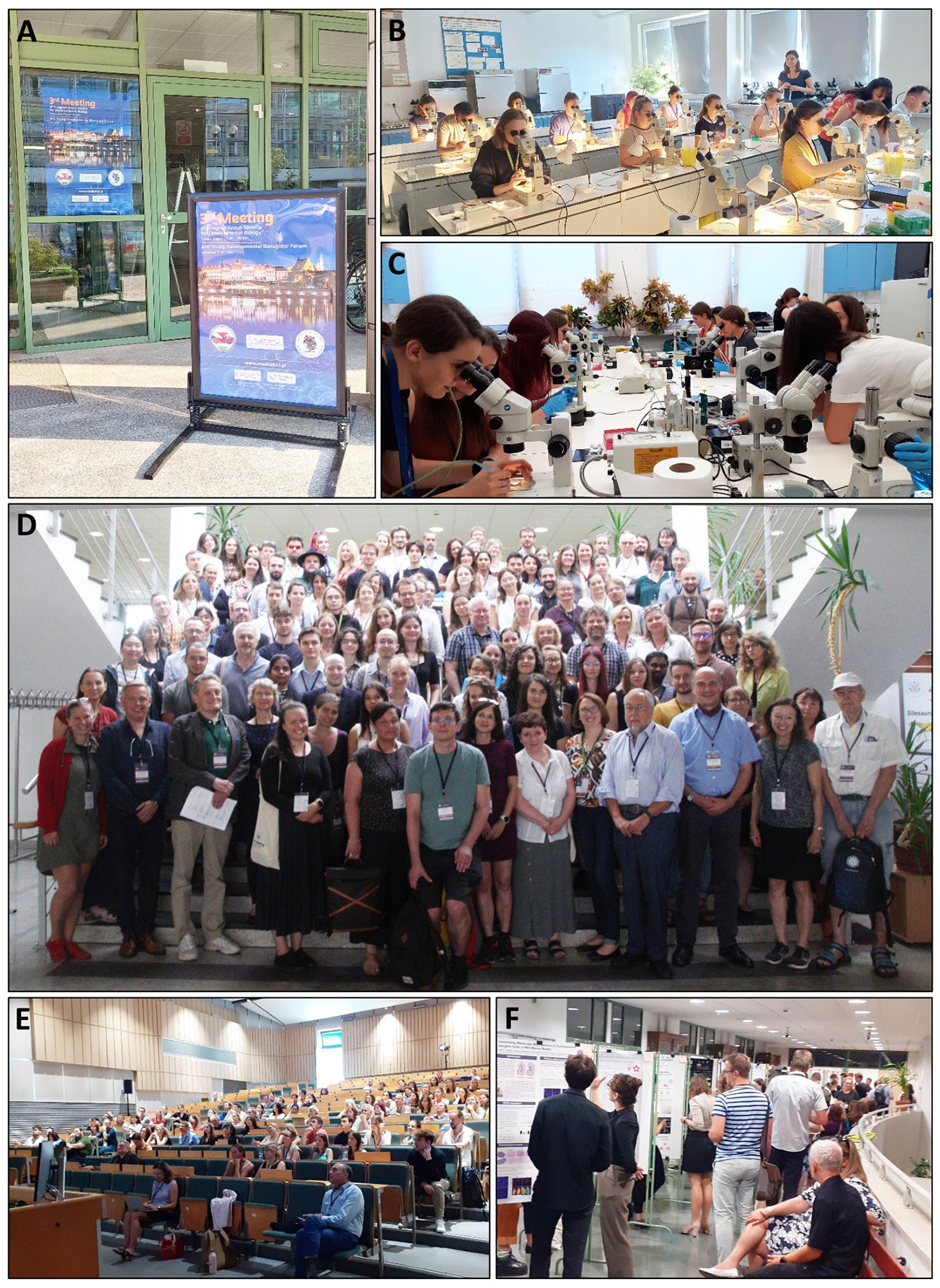
Fig. 2. Overview of the 3rd V4SDB Meeting.
(A) Entrance to the conference venue, Faculty of Biology, University of Warsaw. (B,C) Workshops for young researchers organized during the Young Developmental Biologists’ Forum on September 7th, 2023, and dedicated to (B) chick embryos and (C) mouse oocytes. (D) The conference group photo. There were 160 participants from all over the world. (E) One of the lecture sessions. (F) The poster session.
The main conference was also accompanied by an online outreach event “Developmental Biology for Everyone”. The event was dedicated to secondary and high school students, but welcomed everyone interested in developmental biology. It consisted of two lectures, one in Polish given by Karolina Archacka (University of Warsaw, Poland) on stem cells and the second in English given by Marek Jindra (Biology Center, Czech Academy of Sciences, Czech Republic) on insect metamorphosis.
Organogenesis – the story of cells interacting to form organs
The conference was opened by a lecture session dedicated to organogenesis. An invited speaker, Marianne Bronner (California Institute of Technology, USA) introduced the audience to the cutting-edge research on neural crest cells. The evolutionary development of neural crest cells is unique to vertebrates (Green et al., 2015). These specific stem cells are characterized by multipotency and migratory behavior, and during development, they differentiate into diverse cell types and populate various sites. Marianne showed how the neural crest cells, starting from their induction at the neural plate border until their differentiation into various cell types along the body axis, are defined through the functional characterization of transcriptional and signalling components of neural crest gene regulatory networks. Marianne’s lecture was followed by several short talks covering different aspects of organogenesis. Vladimír Soukup (Charles University, Czech Republic) discussed the role of organizers such as the apical ectodermal ridge (AER) and proposed the existence of such a structure in the outgrowth and axial patterning of the amphibian external gills. Aleksandra Marconi (University of Cambridge, UK) shed light on the genetic basis of species-specific differences in neural crest development in fish. The organogenesis of the vomeronasal organ in snakes, Lacertidae, Iguania, and Gekkota was described in the presentation by Paweł Kaczmarek (University of Silesia, Poland), who also showed substantial organizational differences in the vomeronasal sensory epithelium between snakes and non-ophidian squamates. The next speaker, Peter Fabian (Masaryk University, Czech Republic), defined a novel specialized fibroblast subtype lineage expressing genes of the Phe/Tyr breakdown pathway in zebrafish, suggesting an unexpected local role for Phe/Tyr catabolism outside the liver. The last speaker of this session, Wojciech J. Szlachcic (Adam Mickiewicz University, Poland), revealed how the pancreas follows a precisely orchestrated mechanism of development, in which each pancreatic cell subtype plays a specific role.
Early embryonic development – how the life begins
The session “Early embryonic development” was divided into two parts and covered topics on the early embryo development of different species such as Xenopus, mouse, sheep, wisent, bovine, and axolotl and different experimental approaches used in this broad topic. Early embryogenesis is a complex, precisely controlled process which begins with fertilization and is followed by blastulation and gastrulation leading to the formation of three germ layers: endoderm, ectoderm, and mesoderm (Molè et al., 2020; Płusa and Piliszek, 2020; Solnica-Krezel and Sepich, 2012). In mammals, embryos at the blastocyst stage must implant in the uterus to continue their development. Our adventure with early embryonic development started with the lecture given by invited speaker Michele Boiani (Max Planck Institute for Molecular Medicine, Germany) who discussed the importance of zona pellucida (ZP) in mouse oocytes. Zona pellucida is a glycoprotein coat surrounding mammalian oocytes and until recently, it has been believed that its functions were limited only to the extracellular space where it played a role in mediating sperm binding, preventing polyspermy, and precluding premature embryo-oviductal epithelium interaction. However, Michele shed new light on the ZP functions, indicating that subfraction of ZP proteins operate also in the cytoplasm of mouse oocytes and embryos, where they are necessary during oocyte-to-embryo transition (Israel et al., 2023). Additionally, Michele reported his newest research on the topology of the first embryonic division in mice (Boiani et al., 2023).
The second invited lecture in this session, given by Monika Kaczmarek (Institute of Animal Reproduction and Food Research, Polish Academy of Sciences, Poland), was dedicated to the role of extracellular vesicles (EVs) in embryo-maternal communication during implantation and early pregnancy. Monika demonstrated the presence of EVs, carrying a plethora of miRNAs, in the porcine uterine lumen during early pregnancy. These miRNAs can be internalized by porcine trophoblast cells where they can affect gene expression, stimulate proliferation, and inhibit migration and invasion (Guzewska et al., 2023a; Guzewska et al., 2023b; Szuszkiewicz et al., 2022).
The series of short presentations started with David Drutovic (Institute of Animal Physiology and Genetics, Czech Academy of Sciences, Czech Republic) reporting his results on the regulation of cell cycle progression in mouse embryos by CHK1-CDC25A-CDK1 (Knoblochova et al., 2023). Next, Tereza Toralová (Institute of Animal Physiology and Genetics, Czech Academy of Sciences, Czech Republic) talked about the differences in the expression and degradation of several maternal proteins such as DBF4B, CDC25A, TAB1, CBX5, and PIASy during preimplantation development in various species, concluding that the need for maternal protein degradation is species-specific. In turn, Kateřina Šimková (Institute of Biotechnology, Czech Academy of Sciences, Czech Republic) demonstrated the differences in the localization and degradation of many RNAs of germ cell marker genes during early embryo development between Ambystoma mexicanum (axolotl, order Urodela) and Xenopus (order Anura), which can result from distinct mechanisms of germ cell specification. Štefan Čikoš (Institute of Animal Physiology, Slovak Academy of Sciences, Slovakia) demonstrated that the preimplantation development of mouse embryos is affected by glutamate and gamma-aminobutyric acid (GABA), which can act as signalling molecules (Kovaříková et al., 2023; Špirková et al., 2022). Afterwards, Alexander W. Bruce (University of South Bohemia, Czech Republic) described a new mechanism responsible for the initial spatial segregation of blastomeres in mouse embryos (Gahurova et al., 2023). Next, Roberto de la Fuente (Institute of Genetics and Animal Biotechnology, Polish Academy of Sciences, Poland and University of Manchester, UK) indicated the new role of Runt-related transcription factor 1 (RUNX1), while Margherita Moncada (University of Teramo, Italy) demonstrated the function of adenosine triphosphate (ATP) citrate lyase (ACLY) in the preimplantation development of mouse and sheep embryos, respectively. Agnieszka Bernat (Institute of Genetics and Animal Biotechnology, Polish Academy of Sciences and Intercollegiate Faculty of Biotechnology UG&MUG, Poland) discussed the possibility of the formation of bovine interspecies embryonic chimera with wisent somatic cells, whereas Marek Maleszewski (University of Warsaw, Poland) tackled the problem on when the regulation of cell number occurs in mouse chimeric embryos. Finally, Martin Urbán (Hungarian University of Agriculture and Life Sciences, Hungary) presented his data on the inactivation of X-chromosome during the embryonic development of mouse embryos.
Let’s move on - cellular dynamics and migration in developmental biology
Cell migration is one of the fundamental mechanisms coordinating the establishment and maintenance of the multicellular organism (Trepat et al., 2012). Gastrulation, the process leading to the specification of the three germ layers as well as a multilayered body plan (Solnica-Krezel and Sepich, 2012), is a great model for examining cellular movements. In her invited lecture, Lilianna Solnica-Krężel (Washington University School of Medicine in St. Louis, USA) discussed the relationship between zygotic genome activation and subsequent downregulation of maternally inherited mRNAs and proteins (so-called maternal-to-zygotic transition) and gastrulation. Additionally, she shared her work on pluripotent stem cells-based 2D micropatterned gastruloids (Minn et al., 2020, 2021). Next, the floor was taken by Ann Huysseune (Charles University, Czech Republic) who studied the outer ectodermal layer of sturgeon and proposed periderm homology between the zebrafish and the sturgeon. Marcos Gonzalez Lopez (Masaryk University, Czech Republic) introduced a novel BEE-ST (Bone and tEEth Spatio-Temporal technique) tool, which allows the tracing and quantification of spatiotemporal changes of fully mineralized biological samples in vivo (Gonzalez Lopez et al., 2023). Ewelina Trela (University of Helsinki, Finland, currently University of Warsaw, Poland) and Zuzana Sumbalova Koledova (Masaryk University, Czech Republic) discussed different mechanisms regulating mammary gland development, with a special emphasis on migration-driven cell influx (Trela et al., 2021) and specification of a highly specialized, spatiotemporally restricted subpopulation of fibroblasts unique to puberty, so-called the peri-TEB fibroblasts. The last speaker of this session, Agnieszka Walewska (University of Warsaw, Poland) focused on intracellular cytoplasmic movement in mouse trophectodermal cells and demonstrated that its speed depends on the keratin cytoskeleton and may serve as a valuable and non-invasive biomarker of embryo quality.
Master regulators - genetic and epigenetic control of developmental processes
A fully functional organism, a vertebrate or invertebrate, is built through tightly regulated processes orchestrated by genes. Genes are transcribed into messenger RNAs, which in turn are translated into proteins. Other mechanisms, including epigenetic modifications and non-coding small RNAs, modulate this process by adding additional levels of complexity but also maintaining genomic stability (Hombach and Kretz, 2016). The session on genetic and epigenetic control of developmental processes was opened by invited speaker Petr Svoboda (Institute of Molecular Genetics, Czech Academy of Sciences, Czech Republic), who discussed the role and function of small interfering RNAs (siRNAs) and miRNAs in mammalian female germline (Flemr et al., 2013). The next speaker, Joanna W. Jachowicz (Institute of Molecular Biotechnology, Austrian Academy of Sciences, Austria), continued on the subject of non-coding RNAs (ncRNAs), demonstrating how, through a spatial amplification mechanism, ncRNA Xist achieves chromosome-wide gene silencing of the X chromosome. Additionally, she showed that the newly developed method RNA-DNA SPRITE could be utilized for 3D nuclear localization of RNAs and identification of their genomic targets at the genome-wide scale (Quinodoz et al., 2021). Denisa Jansova (Institute of Animal Physiology and Genetics, Czech Academy of Sciences, Czech Republic) focused on the spatial and temporal translation of specific mRNAs and showed that cyclin B1 and Mos mRNAs localize to the cytoplasm of fully-grown germinal vesicle (GV) oocytes and form cloud-like structures. Next, Adam Kłosin, (Nencki Institute of Experimental Biology, Polish Academy of Sciences, Poland) shared his work on the spatial organization of transcription in developing C. elegans and showed dynamic intranuclear condensates forming during embryonic development. DNA damage was discussed by Helena Fulková (Institute of Experimental Medicine, Czech Academy of Sciences, Czech Republic) who demonstrated that oocytes have the machinery to recognize and repair single-strand DNA breaks (SSBs) protecting the early embryo. In the last talk of this session, Máté Varga (ELTE Eötvös Loránd University, Hungary) showed that a specific regulatory mechanism exists for uracil levels in a zebrafish embryo.
Stem cells and regeneration - developmental biology is not just about embryos
Stem cells, a population of undifferentiated cells with the ability to self-renewal, can differentiate into various cell types. Due to these properties, stem cells are an increasingly important subject of both basic research and clinical trials, as their therapeutic potential is widely tested in medicine (Hoang et al., 2022). During his invited lecture, Giulio Cossu (University of Manchester, UK and San Raffaele Hospital, Italy) demonstrated that mesoangioblasts, derived from adventitial pericytes of skeletal muscle, may be promising candidates for therapies of muscular dystrophies. Recently, Giulio’s team has developed a new strategy that compensates for the low engraftment of transplanted mesoangioblasts in patients’ muscles by genetically amplifying their dystrophin production (Cossu et al., 2023) and now works on immune-privileged mesoangioblasts as universal donor cells. The next speaker, Radek Sindelka (Institute of Biotechnology, Czech Academy of Sciences, Czech Republic), opened the floor for short, and thematically diverse, talks. He talked about the role of a novel cell population, named Regeneration Initiation Cells (RICs), during tail regeneration in Xenopus laevis embryos (Sindelka et al., 2023). The second short talk was given by Jan Krivanek (Masaryk University, Czech Republic), who works on continuously growing mouse teeth and described the stem cell niche as a microenvironment with high adaptability to changing environmental conditions (Krivanek et al., 2020). Tibor Kovács (ELTE Eötvös Loránd University, Hungary) gave the talk about stem cell regeneration in the Drosophila male germline, which is mediated by JAK-STAT-upregulated mitophagy. During the next talk, Vladimír Krylov (Charles University, Czech Republic) described the regenerative potential of Xenopus tropicalis Sertoli cell progenitors (XtiSCs). Last, but not least, Aneta Skoniecka (Medical University of Gdansk, Poland) presented her results on the effects of neoadjuvant therapy (radiotherapy or chemotherapy) on the mobility of fibroblasts co-cultured with autologous adipose-derived stromal cells (ASCs).
How to study development and disease? The need for in vitro models
In vitro models allow the study of a wide range of in vivo biological processes such as tissue renewal, stem cell/niche functions, embryo development, and tissue responses to drugs, mutation, or damage. Although they cannot fully mimic the natural environment, they have been greatly improved in recent years and are widely used as disease models (myocardial ischemic injury (Chen and Vunjak-Novakovic, 2018), Alzheimer’s disease (Sharma et al., 2021), cancer (Katt et al., 2016)) and in developmental biology (El Azhar and Sonnen, 2021; Fatehullah et al., 2016; Kim et al., 2023). Therefore, the last session of the V4SDB meeting was dedicated to in vitro models and their role in research on development and disease. The session started with an invited lecture by Alfonso Martinez Arias (Pompeu Fabra University, Spain) focusing on gastruloids, aggregates of ES cells that under defined culture conditions undergo controlled proliferation, symmetry breaking, and germ layer organization. Alfonso discussed how gastruloids can change our understanding of developmental events in mammals (Arias et al., 2022; Beccari et al., 2018). A series of short talks began with Maria Anna Ciemerych-Litwinienko (University of Warsaw, Poland), who discussed various experimental approaches in studying myogenic differentiation of pluripotent stem cells (Mierzejewski et al., 2023; Świerczek-Lasek et al., 2022). On the other hand, Józef Dulak (Jagiellonian University, Poland) demonstrated changes in iron metabolism in human Duchenne muscular dystrophy (DMD) cardiomyocytes and possible DMD cardiomyopathy relief strategies. Next, Lilla Kókity (Biological Research Centre and University of Szeged, Hungary) focused on the RYBP (RING1 and YY1 Binding Protein) protein, which turned out to be a key regulator of gene expression and endothelial and cardiac lineage commitment (Henry et al., 2023). Then Paulina Gębala (Mossakowski Medical Research Institute, Polish Academy of Sciences, Poland) demonstrated that the autophagy process in the neonatal rat glial cells is actively ongoing during neurodevelopment and that its efficiency can be disturbed by pathophysiological conditions. The last talk in this session was given by Katarzyna Błaszczyk (Adam Mickiewicz University, Poland), who discussed the inhibitory effect of SPOCK2 protein on the proliferation and functioning of human early pancreatic β-cells.
In summary, the third V4SDB meeting gave the participants a unique insight into the most interesting and novel research conducted in the field of developmental biology. A wide scope of themes and model organisms was covered, emphasizing the diversity of developmental biology and clearly illustrating that this discipline still hides many undiscovered phenomena. Fruitful discussions over the coffee breaks and the superb gala dinner resulted in new friendships and collaborations. The next meeting will be held in Slovakia in 2025 where hopefully once more we will be able to celebrate developmental biology in Central Europe.
References
Arias A. M., Marikawa Y., Moris N. (2022). Gastruloids: Pluripotent stem cell models of mammalian gastrulation and embryo engineering. Developmental Biology 488: 35-46.
Beccari L., Moris N., Girgin M., Turner D. A., Baillie-Johnson P., Cossy A.C., Lutolf M. P., Duboule D., Arias A. M. (2018). Multi-axial self-organization properties of mouse embryonic stem cells into gastruloids. Nature 562: 272-276.
Boiani M., Fuellen G., Halabian R., Makalowski W., Nolte T., Suzuki Y., Israel S. (2023). O-179 Fertilization topologies that occur seldom or not at all in nature give rise to distinct functional classes of mouse embryos. Human Reproduction 38: dead093.220.
Chen T., Vunjak-Novakovic G. (2018). In Vitro Models of Ischemia-Reperfusion Injury. Regenerative Engineering and Translational Medicine 4: 142-153.
Cossu G., Tonlorenzi R., Brunelli S., Sampaolesi M., Messina G., Azzoni E., Benedetti S., Biressi S., Bonfanti C., Bragg L., Camps J., Cappellari O., Cassano M., Ciceri F., Coletta M., Covarello D., Crippa S., Cusella-De Angelis M. G., De Angelis L., Dellavalle A., Diaz-Manera J., Galli D., Galli F., Gargioli C., Gerli M. F. M., Giacomazzi G., Galvez B. G., Hoshiya H., Guttinger M., Innocenzi A., Minasi M. G., Perani L., Previtali S. C., Quattrocelli M., Ragazzi M., Roostalu U., Rossi G., Scardigli R., Sirabella D., Tedesco F. S., Torrente Y., Ugarte G. (2023). Mesoangioblasts at 20: From the embryonic aorta to the patient bed. Frontiers in Genetics 13: 1056114.
el Azhar Y., Sonnen K. F. (2021). Development in a Dish—In Vitro Models of Mammalian Embryonic Development. Frontiers in Cell and Developmental Biology 9: 655993.
Fatehullah A., Tan S. H., Barker N. (2016). Organoids as an in vitro model of human development and disease. Nature Cell Biology 18: 246-254.
Flemr M., Malik R., Franke V., Nejepinska J., Sedlacek R., Vlahovicek K., Svoboda P. (2013). A Retrotransposon-Driven Dicer Isoform Directs Endogenous Small Interfering RNA Production in Mouse Oocytes. Cell 155: 807-816.
Gahurova L., Tomankova J., Cerna P., Bora P., Kubickova M., Virnicchi G., Kovacovicova K., Potesil D., Hruska P., Zdrahal Z., Anger M., Susor A., Bruce A. W. (2023). Spatial positioning of preimplantation mouse embryo cells is regulated by mTORC1 and m 7 G-cap-dependent translation at the 8- to 16-cell transition . Open Biology 13: 230081.
Gonzalez Lopez M., Huteckova B., Lavicky J., Zezula N., Rakultsev V., Fridrichova V., Tuaima H., Nottmeier C., Petersen J., Kavkova M., Zikmund T., Kaiser J., Lav R., Star H., Bryja V., Henyš P., Vořechovský M., Tucker A. S., Harnos J., Buchtova M., Krivanek J. (2023). Spatiotemporal monitoring of hard tissue development reveals unknown features of tooth and bone development. Science Advances 9: eadi0482.
Green S. A., Simoes-Costa M., Bronner M. E. (2015). Evolution of vertebrates as viewed from the crest. Nature 520: 474-482.
Guzewska M. M., Myszczynski K., Heifetz Y., Kaczmarek M. M. (2023a). Embryonic signals mediate extracellular vesicle biogenesis and trafficking at the embryo–maternal interface. Cell Communication and Signaling 21: 210.
Guzewska M. M., Witek K. J., Karnas E., Rawski M., Zuba‐Surma E., Kaczmarek M. M. (2023b). miR-125b-5p impacts extracellular vesicle biogenesis, trafficking, and EV subpopulation release in the porcine trophoblast by regulating ESCRT-dependent pathway. The FASEB Journal 37: e23054.
Henry S., Kokity L., Pirity M. K. (2023). Polycomb protein RYBP activates transcription factor Plagl1 during in vitro cardiac differentiation of mouse embryonic stem cells. Open Biology 13: 220305.
Hoang D. M., Pham P. T., Bach T. Q., Ngo A. T. L., Nguyen Q. T., Phan T. T. K., Nguyen G. H., Le P. T. T., Hoang V. T., Forsyth N. R., Heke M., Nguyen L. T. (2022). Stem cell-based therapy for human diseases. Signal Transduction and Targeted Therapy 7: 272.
Hombach S., Kretz M. (2016). Non-coding RNAs: Classification, Biology and Functioning. In Non-coding RNAs in Colorectal Cancer. (Ed. Slaby O., Calin G. A.) Springer International Publishing, Cham.
Israel S., Seyfarth J., Nolte T., Drexler H. C. A., Fuellen G., Boiani M., (2023). Intracellular fraction of zona pellucida protein 3 is required for the oocyte-to-embryo transition in mice. Molecular human reproduction 29: gaad038.
Katt M. E., Placone A. L., Wong A. D., Xu Z. S., Searson P. C. (2016). In Vitro Tumor Models: Advantages, Disadvantages, Variables, and Selecting the Right Platform. Frontiers in Bioengineering and Biotechnology 4: 12.
Kim Y., Kim I., Shin K. (2023). A new era of stem cell and developmental biology: from blastoids to synthetic embryos and beyond. Experimental & Molecular Medicine 55: 2127-2137.
Knoblochova L., Duricek T., Vaskovicova M., Zorzompokou C., Rayova D., Ferencova I., Baran V., Schultz R. M., Hoffmann E. R., Drutovic D. (2023). CHK1-CDC25A-CDK1 regulate cell cycle progression and protect genome integrity in early mouse embryos. EMBO reports 24: e56530.
Kovaříková V., Špirková A., Šefčíková Z., Pisko J., Kalatová L., Koppel J., Fabian D., Čikoš Š (2023). Gamma‐aminobutyric acid (GABA) can affect physiological processes in preimplantation embryos via GABA A and GABA B receptors. Reproductive Medicine and Biology 22: e12528.
Krivanek J., Soldatov R. A., Kastriti M. E., Chontorotzea T., Herdina A. N., Petersen J., Szarowska B., Landova M., Matejova V. K., Holla L. I., Kuchler U., Zdrilic I. V., Vijaykumar A., Balic A., Marangoni P., Klein O. D., Neves V. C. M., Yianni V., Sharpe P. T., Harkany T., Metscher B. D., Bajénoff M., Mina M., Fried K., Kharchenko P. V., Adameyko I. (2020). Dental cell type atlas reveals stem and differentiated cell types in mouse and human teeth. Nature Communications 11: 4816.
Mierzejewski B., Grabowska I., Michalska Z., Zdunczyk K., Zareba F., Irhashava A., Chrzaszcz M., Patrycy M., Streminska W., Janczyk-Ilach K., Koblowska M., Iwanicka-Nowicka R., Gromadka A., Kowalski K., Ciemerych M. A., Brzoska E. (2023). SDF-1 and NOTCH signaling in myogenic cell differentiation: the role of miRNA10a, 425, and 5100. Stem Cell Research & Therapy 14: 204.
Minn K. T., Dietmann S., Waye S. E., Morris S. A., Solnica-Krezel L. (2021). Gene expression dynamics underlying cell fate emergence in 2D micropatterned human embryonic stem cell gastruloids. Stem Cell Reports 16: 1210-1227.
Minn K. T., Fu Y. C., He S., Dietmann S., George S. C., Anastasio M. A., Morris S. A., Solnica-Krezel L. (2020). High-resolution transcriptional and morphogenetic profiling of cells from micropatterned human ESC gastruloid cultures. eLife 9: e59445.
Molè M. A., Weberling A., Zernicka-Goetz M. (2020). Comparative analysis of human and mouse development: From zygote to pre-gastrulation. In Gastrulation: From Embryonic Pattern to Form. Elsevier.
Płusa B., Piliszek A. (2020). Common principles of early mammalian embryo self-organisation. Development 147: dev183079.
Quinodoz S. A., Jachowicz J. W., Bhat P., Ollikainen N., Banerjee A. K., Goronzy I. N., Blanco M. R., Chovanec P., Chow A., Markaki Y., Thai J., Plath K., Guttman M. (2021). RNA promotes the formation of spatial compartments in the nucleus. Cell 184: 5775-5790.e30.
Sindelka R., Abaffy P., Zucha D., Naraine R., Kraus D., Netusil J., Smetana K., Lukas L., Endaya B. B., Neuzil J., Psenicka M., Kubista M., (2023). Characterization of regeneration initiating cells during Xenopus laevis tail regeneration. BioRxiv Preprint: .
Sharma N. S., Karan A., Lee D., Yan Z., Xie J. (2021). Advances in Modeling Alzheimer's Disease In Vitro. Advanced NanoBiomed Research 1: 2100097.
Solnica-Krezel L., Sepich D. S. (2012). Gastrulation: Making and Shaping Germ Layers. Annual Review of Cell and Developmental Biology 28: 687-717.
Špirková A., Kovaříková V., Šefčíková Z., Pisko J., Kšiňanová M., Koppel J., Fabian D., Čikoš Š (2022). Glutamate can act as a signaling molecule in mouse preimplantation embryos. Biology of Reproduction 107: 919-916.
Świerczek-Lasek B., Tolak L., Bijoch L., Stefaniuk M., Szpak P., Kalaszczynska I., Streminska W., Ciemerych M. A., Archacka K. (2022). Comparison of Muscle Regeneration after BMSC-Conditioned Medium, Syngeneic, or Allogeneic BMSC Injection. Cells 11: 2843.
Szuszkiewicz J., Myszczynski K., Reliszko Z. P., Heifetz Y., Kaczmarek M. M. (2022). Early steps of embryo implantation are regulated by exchange of extracellular vesicles between the embryo and the endometrium. The FASEB Journal 36: e22450.
Trela E., Lan Q., Myllymäki S.M., Villeneuve C., Lindström R., Kumar V., Wickström S. A., Mikkola M. L. (2021). Cell influx and contractile actomyosin force drive mammary bud growth and invagination. Journal of Cell Biology 220: e202008062.
Trepat X., Chen Z., Jacobson K., (2012). Cell Migration. Comprehensive Physiology 2: 2369-2392.